Magnetic Resonance Imaging (MRI) uses a strong magnetic field and radio frequency pulses to provide clear and detailed diagnostic images of internal body organs and tissues. MRI is a valuable tool for the diagnosis of a broad range of conditions, including, but not limited to:
 Cancer
 Heart and vascular disease
 Stroke
 Joint and musculoskeletal disorders
MRI allows evaluation of some bodily structures that may not be as visible with other diagnostic imaging methods.
What are some common uses of MRI?
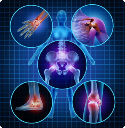
Imaging of the Musculoskeletal System
MRI is often used to study the knee, ankle, foot, shoulder, elbow, wrist, and hand. MRI is also a highly accurate method for evaluating soft tissue structures, such as tendons and ligaments, which can be seen in great detail with this method. Many subtle injuries are easily detected. In addition, MRI is used for the diagnosis of spinal problems, including disc herniation, spinal stenosis, and spinal tumors.
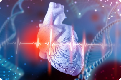
Imaging of the Heart
MRI of the heart, aorta, coronary arteries, and blood vessels is a tool for diagnosing coronary artery disease and other heart problems. Using MRI, doctors can examine the size and thickness of the chambers of the heart and determine the extent of damage caused by a heart attack or heart disease.
Imaging for Cancer and Functional Disorders. Organs of the chest and abdomen, such as the liver, lungs, kidney and other abdominal organs, can be examined in great detail with MRI. This aids in the diagnosis and evaluation of tumors and functional disorders. For early diagnosis of breast cancer, MRI is a supplement to traditional mammography. Furthermore, because no radiation exposure is involved, MRI is often used for examination of the male and female reproductive systems.
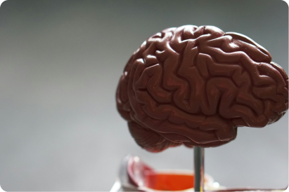
Imaging of the Head and Spine
MRI for neurological/brain imaging and spine studies provides outstanding image quality for diagnosis. The MRI software offers many mode and viewing options including the ability to reconstruct and rotate images to show soft tissue of the brain. MRA studies offer enhanced images of vascular structures.
What should I expect during this exam?

Duration
Depending on how many images are needed, the exam generally takes 20 to 45 minutes. Very detailed studies may take longer.

Positioning
You will be asked to lie down on a sliding table and will be positioned comfortably.

Communication
The technologist and nurse will leave the room for your exam, but you will be able to communicate with them at any time using an intercom.

Contrast Material
Depending on the part of the body being examined, a contrast material may be used to enhance the visibility of certain tissues or blood vessels.

Scanner Noise
You will hear loud tapping or thumping during the exam. Patients may choose either music, earplugs, or both to block out the noise.

Medical Support
In certain circumstances, a friend or family member will be allowed to stay in the room with you during the exam.